An asterisk (*) marks authors the paper states contributed equally as first authors. Other sections list papers newest first.
First-author peer-reviewed publications
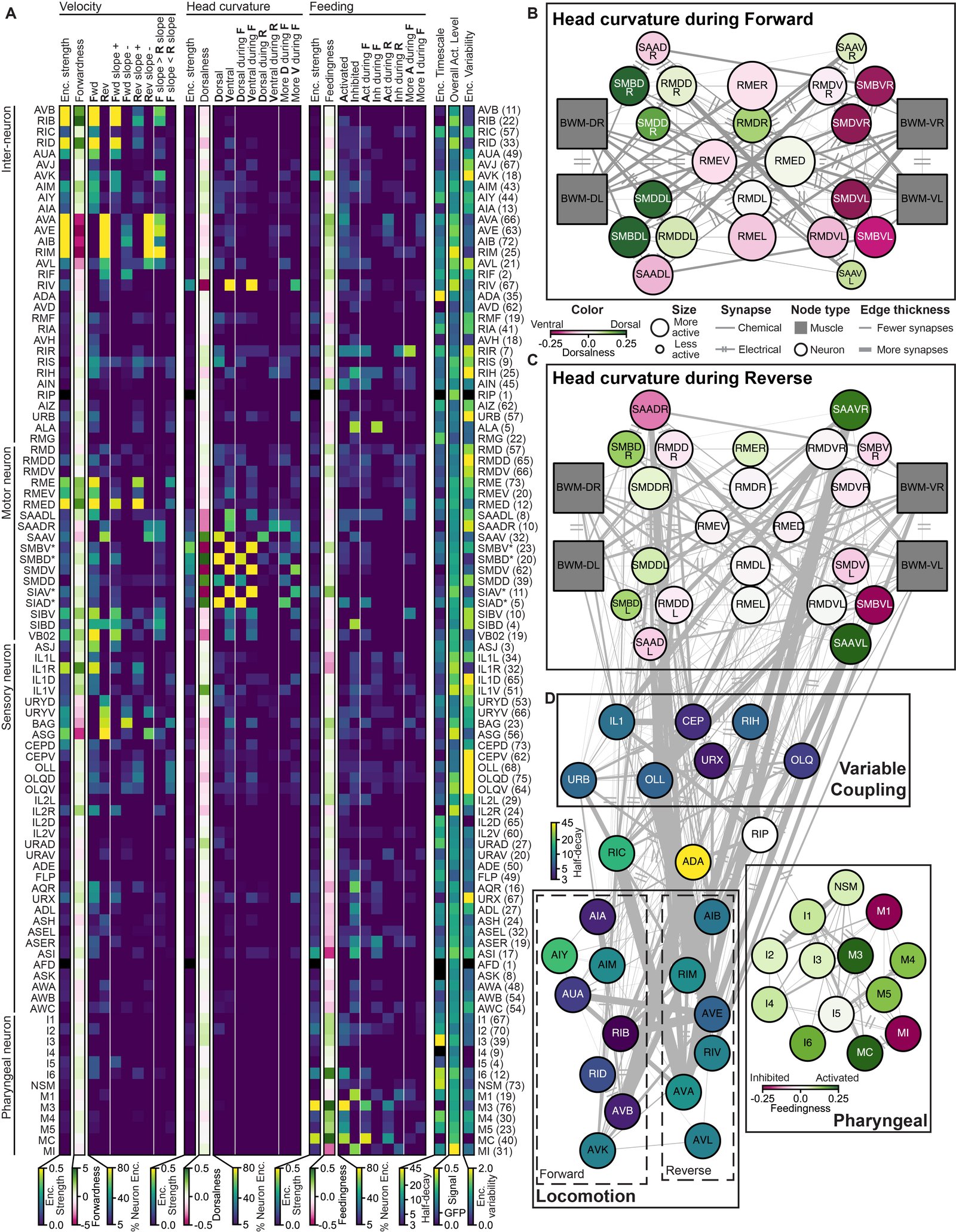
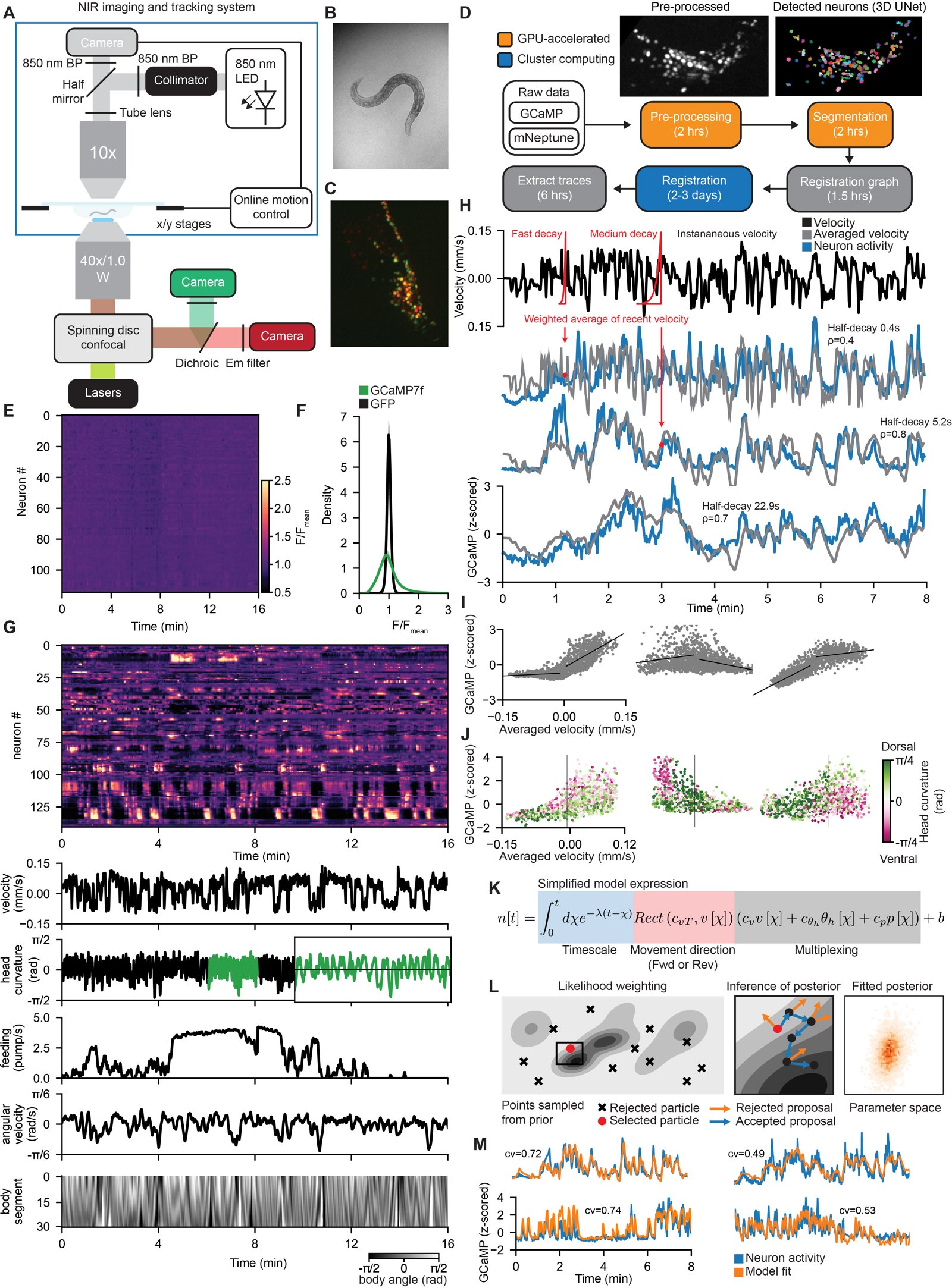
Atanas, A. A.*, Kim, J.*, et al. (2023). Brain-wide representations of behavior spanning multiple timescales and states in C. elegans. Cell 186, 4134–4151.e31. doi:10.1016/j.cell.2023.07.035 · wormwideweb.org
Jungsoo Kim and I, as co-first authors, asked how neurons across an entire brain encode an animal's behavior, and how that encoding changes with the animal's internal state. We built a microscope that tracks a freely moving worm while imaging calcium activity across its head, and recorded brain-wide activity together with its velocity, head curvature, feeding and body posture. Our image-analysis pipeline (ANTSUN) turned these recordings into neural traces, and in 40 of the recordings we used NeuroPAL multicolor labeling to identify which defined neuron class each recorded neuron belonged to.
To explain each neuron's activity, we built the C. elegans Probabilistic Neural Encoding Model (CePNEM), which fits how a neuron's activity depends on these behaviors, including the timescale over which it integrates recent actions. Because CePNEM infers a full posterior distribution over these parameters rather than a single best fit, we could say with stated confidence which behaviors each neuron encodes. Many neuron classes encode several behaviors at once, some track the animal's current motion while others integrate its actions over tens of seconds, and when a heat stimulus shifts the animal's behavioral state, many neurons change how they encode behavior. We combined these results into an atlas of how the neuron classes of the C. elegans connectome encode behavior, and released the recordings at WormWideWeb, where anyone can browse and download them.
Figure 4: an atlas of how each identified neuron class encodes velocity, head curvature and feeding, mapped onto the connectome.Figure 1: the tracking microscope and analysis pipeline, an example recording, neurons that encode velocity over different timescales, and the CePNEM model.
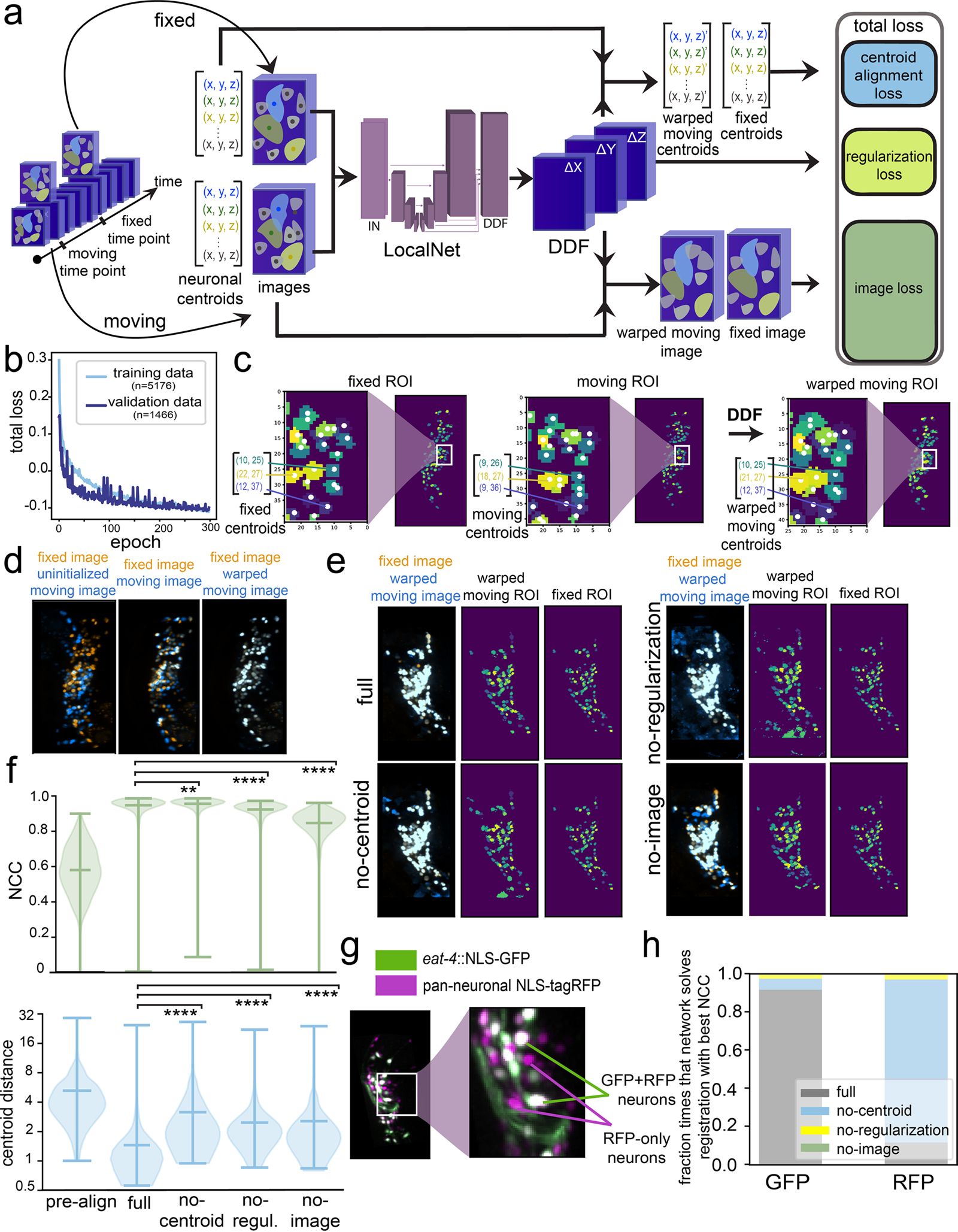
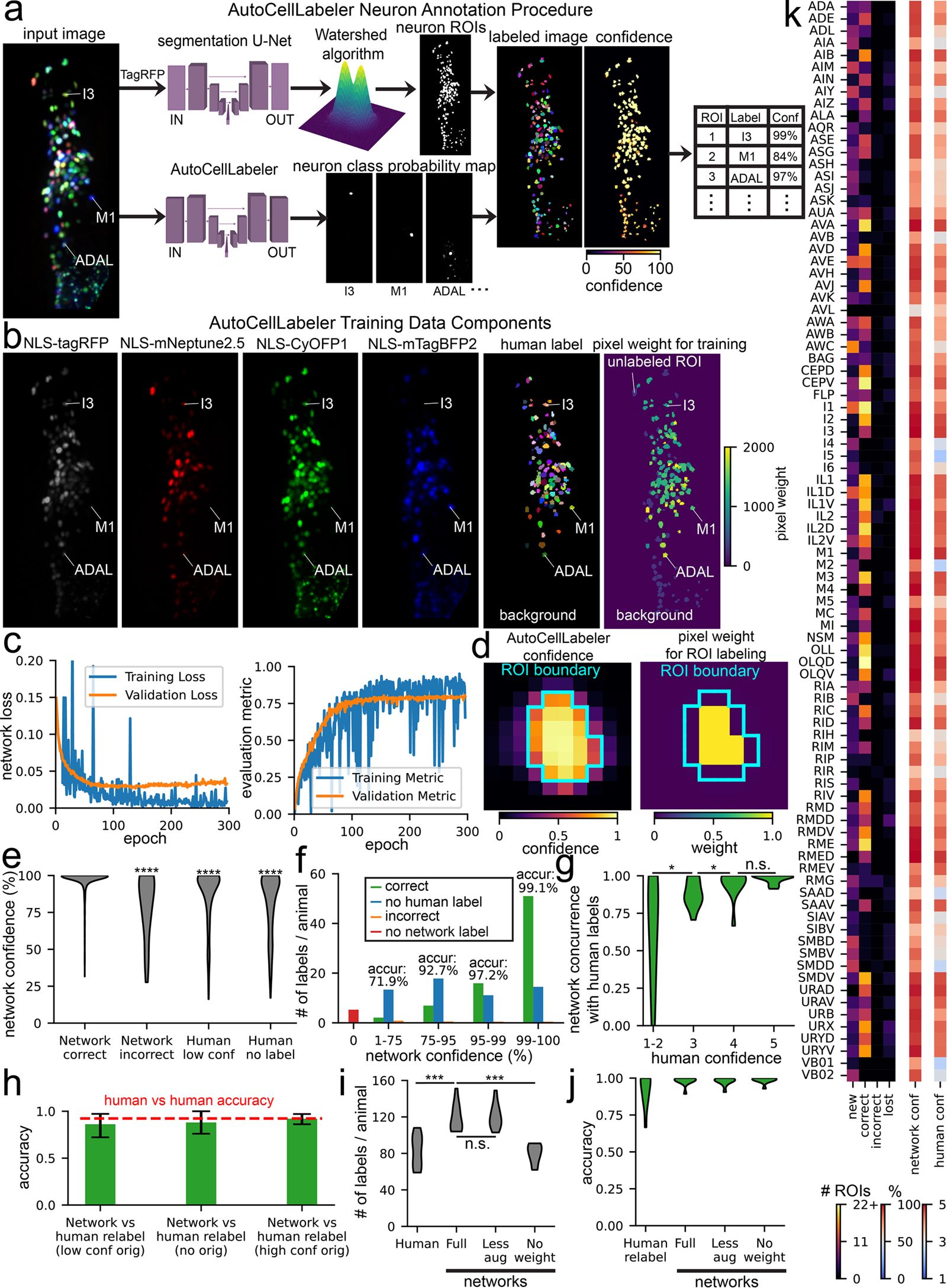
Atanas, A. A., et al. (2026). Deep neural networks to register and annotate cells in moving and deforming nervous systems. eLife 14:RP108159. doi:10.7554/eLife.108159.2
Recording neurons in a moving animal requires two things: following each neuron as the brain bends and deforms, and knowing which neuron is which. This paper presents three deep neural networks that automate both. BrainAlignNet aligns pairs of images of the bending C. elegans head with single-pixel accuracy, using both the images and the positions of neuron centers to train it; within our ANTSUN pipeline it links neurons across time with 99.6% accuracy, and it also aligns neurons in the jellyfish Clytia hemisphaerica, an animal with a very different body plan.
AutoCellLabeler reads the multicolor NeuroPAL fluorescence and labels more than 100 neuronal cell types per animal with 98% accuracy, beating individual human labelers by learning from many manually labeled datasets. A third network, CellDiscoveryNet, compares imaging data across many animals to discover and label cell types without any human labels, and matches trained human labelers. I am the first author; my contributions included conceptualization, software, analysis and writing.
Figure 1: how BrainAlignNet is trained, and its non-rigid registration of the bending head.Figure 4: AutoCellLabeler annotates more than 100 neuronal cell types from multicolor fluorescence.
Kramer, T. S., Wan, F. K., Pugliese, S. M., Atanas, A. A., et al. (2026). Neural sequences underlying directed turning in Caenorhabditis elegans. Nature Neuroscience 29, 1408–1424. doi:10.1038/s41593-026-02257-5
Using whole-brain calcium imaging and cell-specific perturbations, this study traces error-correcting turns during olfactory navigation in the worm to neural sequences, in which defined neurons activate in a stereotyped order during each turn. Neurons in the sequence respond to odor cues, anticipate turn direction and drive movement, and the neuromodulator tyramine coordinates these dynamics; I was one of the authors who developed the software.
Pradhan, S.*, Madan, G. K.*, Kang, D., Bueno, E., Atanas, A. A., et al. (2025). Pathogen infection induces sickness behaviors through neuromodulators linked to stress and satiety in C. elegans. Nature Communications 16, 3200. doi:10.1038/s41467-025-58478-y
Worms infected with the bacterium Pseudomonas aeruginosa PA14 first reduce feeding, then show reversible bouts of quiescence, and eventually die; this study identifies neurons and neuropeptides behind these changes, including the ALA neuron and a brain-wide imaging screen that implicated the ASI neuron and DAF-7/TGF-beta. The results suggest that a common set of neuromodulators is recruited across physiological states, acting from different neural sources and in different combinations; I contributed to the software.
Dag, U.*, Nwabudike, I.*, Kang, D.*, Gomes, M. A., Kim, J., Atanas, A. A., et al. (2023). Dissecting the functional organization of the C. elegans serotonergic system at whole-brain scale. Cell 186, 2574–2592.e20. doi:10.1016/j.cell.2023.04.023
This study examines how serotonin release alters brain-wide activity to induce foraging behaviors such as slow locomotion and increased feeding, identifying core receptors for slow locomotion and showing that the receptor SER-4 drives behavioral responses to sudden increases in release while MOD-1 drives responses to persistent release. Mapping all sites of serotonin receptor expression in the connectome, together with synaptic connectivity, helps predict which neurons show serotonin-associated activity; I contributed to methodology and software.
Kuraszkiewicz, J., Wilkes, B. J., Atanas, A., et al. (2021). Beyond Simple AGN Unification with Chandra-observed 3CRR Sources at 0.5 < z < 1. The Astrophysical Journal 913, 134. doi:10.3847/1538-4357/abf3c0
We report Chandra X-ray observations of a complete, orientation-unbiased sample of 44 radio-selected active galactic nuclei (AGN) at redshift z between 0.5 and 1. Obscuration generally increases with inclination, as simple unification predicts, but some narrow-line radio galaxies at intermediate inclinations are less obscured than expected, suggesting that the Eddington ratio (luminosity relative to the Eddington limit) matters as well as orientation.
Dai, Y. S., Wilkes, B. J., Bergeron, J., Kuraszkiewicz, J., Omont, A., Atanas, A., & Teplitz, H. I. (2018). Is there a relationship between AGN and star formation in IR-bright AGNs? Monthly Notices of the Royal Astronomical Society 478, 4238–4254. doi:10.1093/mnras/sty1341
This paper measures the relationship between AGN luminosity and star formation rate in 323 far-infrared-detected AGN at redshifts from 0.2 to 2.5, and recommends far-infrared luminosity as a more reliable star formation estimator because AGN contribute on average about 20% of total infrared luminosity. The correlation is significant, and the ratio of star formation rate to black hole accretion rate shows no dependence on redshift or mass, consistent with both processes depending on a common gas supply.
Veliz-Cuba, A., Hirning, A. J., Atanas, A. A., et al. (2015). Sources of Variability in a Synthetic Gene Oscillator. PLOS Computational Biology 11, e1004674. doi:10.1371/journal.pcbi.1004674
Using microfluidic devices and time-lapse fluorescence microscopy to track oscillations across cell lineages, this study found high cell-to-cell variability in amplitude while sister cells stayed strongly correlated for many minutes after division. A computational model tuned to these noise sources reproduced the observations and predicted outcomes under new conditions; I was one of the authors who analyzed the data.
Conference proceedings
Prabhakar, V., Islam, M. A., Atanas, A., et al. (2026). OmniScience: A Domain-Specialized LLM for Scientific Reasoning and Discovery. Proceedings of AI for Accelerated Research Symposium, EPiC Series in Technology, vol. 3, 36–49. doi:10.29007/5h18 · arXiv:2503.17604
This paper describes a large language model (LLM) for general science, built through pretraining on scientific literature, instruction tuning, and reasoning-based knowledge distillation, and applied in a battery agent that ranks molecules as candidate electrolyte solvents or additives. The authors report that it is competitive with state-of-the-art reasoning models on the GPQA Diamond and battery benchmarks, and that the pretraining and distillation steps were critical to that performance.
Preprints
Atanas, A., & Liu, K. (2025). A Modular Dataset to Demonstrate LLM Abstraction Capability. arXiv:2503.17645. Preprint.
We introduce ArrangementPuzzle, a puzzle dataset with structured solutions and automated checking of each reasoning step. A classifier trained on LLM activations predicted whether a reasoning step was correct with over 80% accuracy, with the strongest representations in the middle-late layers of the transformer, and further analysis found that these layers distinguish logical from semantic equivalence.
Theses and short reports
Atanas, A. (2023). Brain-wide representations of behavior spanning multiple timescales and states in C. elegans. PhD thesis, Massachusetts Institute of Technology. MIT DSpace
My doctoral thesis in the Computational and Systems Biology Program, advised by Steven Flavell. It covers the same work as the 2023 Cell paper above: brain-wide recordings of freely moving worms, probabilistic models of how each neuron encodes behavior, and how that encoding changes with behavioral state.
Atanas, A. A. (2017). Evolutionary Graph Theory of Biased Sexual Reproduction. Bachelor's thesis, Harvard College. Harvard DASH repository
I proposed a family of models of a mutant strain spreading through a graph-structured population by biasing sexual reproduction in its favor, with possible application to gene drives (genetic elements that bias their own inheritance). The thesis gives analytical results, including the probability that the mutant takes over (its fixation probability) on large complete graphs with a perfect gene drive, along with a simulator; a stated limitation is that the models have no gender, so any individual can mate with any neighbor.
Faesi, C., Bieryla, A., Berger, E., Atanas, A., et al. (2014). Spectroscopic Classification of PSN J07562525+2700488. The Astronomer's Telegram 5993.
A short report from an observing trip in a Harvard undergraduate astronomy course: an optical spectrum taken with the 1.5-meter telescope at the Fred Lawrence Whipple Observatory matched a normal type Ia supernova about 9 days after maximum light.